


|
Renal medical emergencies Nephrology (Kidney Disease) |
|
What are the components of the human urinary system? Can you name the components of the human urinary system? 2 kidneys 2 ureters 1 urinary bladder 1 urethra 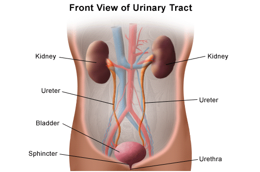 What are the components of the human kidney? Can you name the components of the human kidney? Renal capsule Renal cortex Renal medulla Renal pelvis Calyx: major calyx and minor calyx Nephron Medullary pyramids Ureter Renal arteries Renal veins 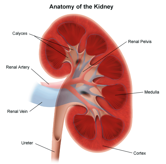 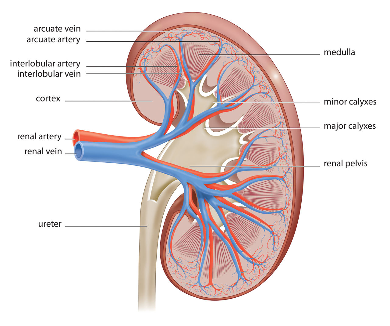 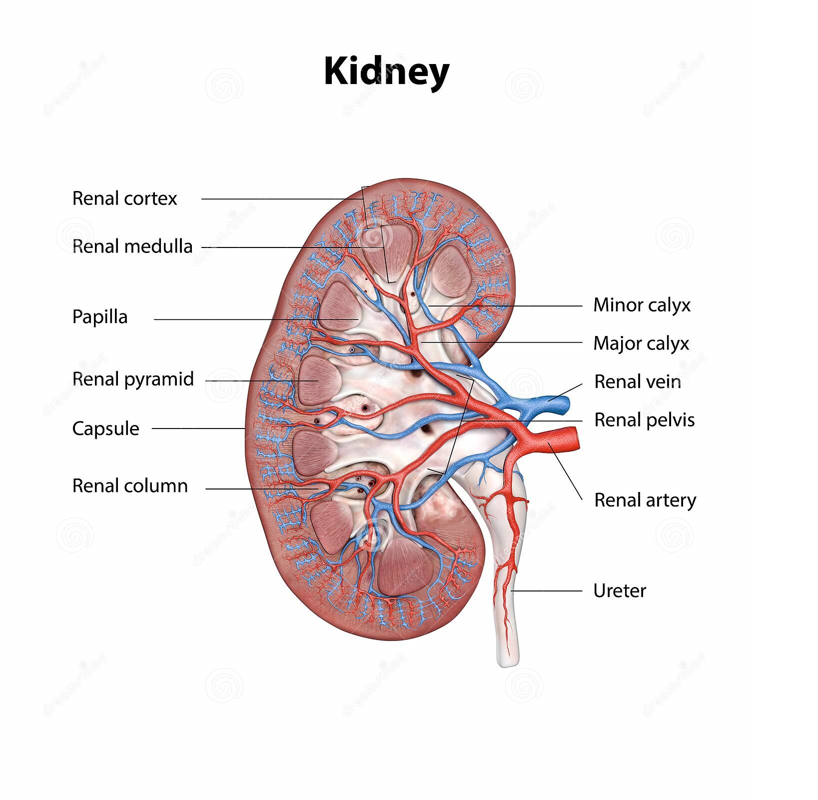
How does the urinary system work? The urinary system's function is to filter blood and create urine as a waste by-product. The organs of the urinary system include the kidneys, renal pelvis, ureters, bladder and urethra. 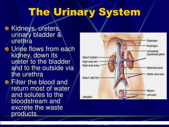
 What is the Nephron? How does a nephron filter blood? What are the two regions of the nephron? Where are the nephrons located? What is the main function of a nephron? What are the two main parts of a nephron structure? What is the cup-shaped structure surrounding the renal corpuscle called? Why Juxtamedullary nephrons are more important in osmoregulation? In this way, how do nephrons maintain homeostasis? Also, do we have more cortical or Juxtamedullary nephrons? Why do kidneys act as both Osmoregulatory and excretory organs? Why is homeostasis important? What is the renal corpuscle? What is the renal tubule? What is the loop of Henle? What are the 2 nephron types? What is the cortical neprhon? What percentage of nephrons are cortical? What is the Nephron? The nephron is the basic functional and structural unit of the kidney, and each human kidney contains from 800,000 to one million of these units. They are responsible for maintaining the concentrations of water and soluble substances in the blood and regulating blood volume, blood pressure, and the blood's pH or acidity. This structure works by filtering the blood, reabsorbing nutrients, and excreting excess water and waste as urine. There are two types of nephrons, distinguished by their location in the kidney. Cortical nephrons are located in the renal cortex on the outside of the organ, while juxtamedullary nephrons are located deeper in the kidney, in the renal medulla. Each nephron is made up of a renal corpuscle and a renal tubule. The renal corpuscle provides the initial filtering component, while the renal tubule is responsible for reabsorption. The corpuscle is composed of the glomerulus and Bowman's capsule. The glomerulus is a bundle of capillaries, or small, permeable blood vessels, through which oxygenated blood enters the kidneys. Excess water and waste products are collected in Bowman's capsule, which houses the glomerulus, and the rest of the blood rejoins the main bloodstream. The renal tubule consists of the proximal tubule, the loop of Henle, and the distal convoluted tubule. Each portion is responsible for a different part of reabsorption. About two-thirds of the filtered salt and water from the renal corpuscle, along with all of the filtered organic solutes, are reabsorbed in the proximal tubule. The loop of Henle has two major portions: the descending limb and the ascending limb. The former is water-permeable, but impermeable to salt, while the later is impermeable to water. Water is removed from the tubule fluid as it passes through the descending limb of the loop of Henle, while sodium is pumped out of the fluid as it passes through the ascending limb. The distal convoluted tubule is controlled by hormones from the endocrine system, causing it to reabsorb or excrete certain nutrients as required for the body's needs. It also regulates blood pH. After reabsorption is complete, the remaining filtrate passes out of the nephron and into the collecting duct system, which collects urine before it is excreted. Urine leaves the collecting ducts through the renal papillae, passing into the renal calyxes, then the renal pelvis, and finally entering the bladder by way of the ureter. How does a nephron filter blood? A nephron consists of a filter called glomerulus and a tubule. The glomerulus filters the fluid and waste products holding back the blood cells and large molecules, especially proteins. What are the two regions of the nephron? Glomerulus, and Renal Tubule. Nephron Sections Glomerulus The glomerulus is a small, spherical structure at the center of which lies a tuft of glomerular capillaries surrounded by the membranous Bowman's Capsule. The Bowman's Capsule is continuous with the proximal tubule and collects any fluid filtering through the glomerular capillaries and directs it into the remainder of the nephron. Proximal Tubule: The proximal tubule lies within the renal cortex and is the section of nephron immediately after the glomerulus. The entire proximal tubule possesses a distinct physiological function and charts a convoluted, twisting path and is often termed the "Proximal Convoluted Tubule". Loop of Henle: The loop of Henle is the section of nephron lying after the proximal tubule and dips down into the renal medulla. Henle's loop displays variable thickness with the entire descending arm and the lower portion of the ascending arm being quite thin, together known as the Thin Loop of Henle. In contrast, the upper portion of the ascending arm is thick and is known as the Thick Ascending Loop of Henle. As suggested by their distinct anatomical and histological appearance the thin and thick segments of Henle's loop display different functionalities. At the end of the thick ascending loop of Henle there is a blister of tissue known as the Macula Densa which is part of the Juxtaglomerular Apparatus. Distal Tubule: The distal tubule follows the loop of Henle and lies within the renal cortex. Like the proximal tubule, the distal tubule charts a convoluted, twisting course and is sometimes termed the Distal Convoluted Tubule. The early distal tubule possesses a distinct functionality than its late portion. The late distal tubule functions in concert with the collecting duct and thus these segments are often termed the Late Distal Tubule and Collecting Duct. Collecting Duct The collecting duct lies after the distal tubule and dips back into the renal medulla. The end of the collecting duct exists at the renal papillae and any remaining nephronic fluid (now considered urine) into the minor calyces. As mentioned, the collecting duct and the late distal tubule share a similar function and are thus grouped under the heading of Late Distal Tubule and Collecting Duct. 
STEP 1: BOWMAN’S CAPSULE Blood pressure in the small capillaries at one end of the nephron (called the glomerulus) pushes fluid into a sack called Bowman’s capsule. Red blood cells and larger proteins are too large to pass from the glomerulus into Bowman’s capsule, but water, ions, and amino acids are small enough to pass through. This mixture of water, ions, amino acids, and other small molecules is called the filtrate. This filtrate is further modified as it travels through the rest of the nephron. STEP 2: PROXIMAL CONVOLUTED TUBULE The filtrate travels from Bowman’s capsule into the proximal convoluted tubule. The goal of this step is to reabsorb (in other words, not excrete) important nutrients including amino acids, glucose, and vitamins that are still present in the filtrate. These are transported out of the nephron into the interstitial space and are reabsorbed by capillaries that run next to the proximal convoluted tubule. In addition to saving important nutrients by transporting them out of the filtrate (reabsorption), waste products, including ammonia and urea, are transported into the filtrate for excretion. STEP 3: DESCENDING LIMB OF THE LOOP OF HENLE The descending limb is only permeable to water. As the Loop of Henle descends from the cortex into the medulla, the concentration of salts in the interstitium increases (you will learn why that happens in the next step). As a result, the medulla is hypertonic to the filtrate, so water will diffuse out of the filtrate on the descending part of the loop, and urine concentration will increase. STEP 4: ASCENDING LIMB OF THE LOOP OF HENLE The ascending limb is only permeable to salts, not water. In the ascending limb, salts (, etc) leave the filtrate using both passive and active diffusion. This is what causes the renal medulla to have a high salt concentration and therefore be hypertonic to the filtrate. The filtrate dilutes on the way up the ascending limb. At the top of the ascending limb, is a thick section called the diluting segment. This section is thick because it contains many mitochondria required to actively pump out salt from the hypotonic filtrate to the more hypertonic blood! STEP 5: DISTAL CONVOLUTED TUBULE You can distinguish this from the proximal convoluted tubule, which is closer to Bowman’s Capsule, because distal means further away (think distance). Functionally it does many of the same things as the proximal convoluted tubule; waste products (ammonia and urea) are transported into the filtrate for excretion, while calcium and sodium continue to be reabsorbed (via active transport out of the nephron into the interstitium). Because water generally follows salt (through osmosis), water also leaves the nephron, which further dilutes the filtrate. STEP 6: COLLECTING DUCTS At the end of the distal convoluted tubule, the filtrate empties into collecting ducts, where it combines with filtrate from other nephrons. Collecting ducts move back into the medulla, which is salty, so more water leaves through passive diffusion. The collecting duct is important because it has variable permeability that is controlled in part by the hormones aldosterone (which increases permeability of the duct to water by opening aquaporins) and antidiuretic hormone (which increases Na/K pump activity), both of which result in a more concentrated filtrate. You may have noticed an organizing principle during this review; in general, the horizontal portions of the nephron function to keep what the body needs and eliminate what it does not need, while the vertical portions of the nephron function to control the quantity and concentration of the filtrate, mostly by controlling the movement of water in and out of the filtrate. Good luck as you work to master this important content! Where are the nephrons located?  The nephrons are located in the cortex and medulla of the kidney. The cortex contains the renal corpuscle, distal convoluted tubule and proximal convoluted tubule. Whereas, the medulla contains the loop of Henle and collecting ducts. What is the main function of a nephron? A nephron is the structural and functional unit of the kidney. It regulates the concentration of water and minerals such as sodium by filtering the blood and reabsorbing the important nutrients. What are the two main parts of a nephron structure? Glomerulus, and Renal Tubule. What is the cup-shaped structure surrounding the renal corpuscle called? The cup-shaped structure surrounding the renal corpuscle is known as the Bowman’s capsule or glomerulus that helps in blood filtration. Why Juxtamedullary nephrons are more important in osmoregulation? Juxtamedullary nephrons, unlike those farther away from the renal medulla, have long nephron loops than extend deep into the medulla. They make the greatest contribution to the osmotic gradient of the medulla, and it's this gradient that enables the kidneys to conserve water and secrete hypertonic urine. In this way, how do nephrons maintain homeostasis? The kidneys remove waste products from metabolism such as urea, uric acid, and creatinine by producing and secreting urine. Urine may also contain sulfate and phenol waste and excess sodium, potassium, and chloride ions. The kidneys help maintain homeostasis by regulating the concentration and volume of body fluids. Beside above, what is the difference between the cortical and Juxtamedullary nephrons? Juxtamedullary nephron is a microscopic structural and functional unit of the kidney with a long loop of Henle. Cortical nephrons have a short loop of Henle, which penetrates only the outer renal medulla. Juxtamedullary nephrons have a long loop of Henle extending deep into the renal medulla. Also, do we have more cortical or Juxtamedullary nephrons? The relative number of cortical and juxtamedullary nephrons and the lengths of their loops of Henle determine the ability of the kidney to concentrate the urine. In humans, about 85% of the nephrons are cortical nephrons and about 15% are juxtamedullary nephrons. Why do kidneys act as both Osmoregulatory and excretory organs? The kidneys are the main osmoregulatory organs in mammalian systems; they function to filter blood and maintain the osmolarity of body fluids at 300 mOsm. The nephron filters and exchanges water and solutes with two sets of blood vessels and the tissue fluid in the kidneys. Why is homeostasis important? Cells depend on the body environment to live and function. Homeostasis keeps the body environment under control and keeps the conditions right for cells to live and function. Without the right body conditions, certain processes (eg osmosis) and proteins (eg enzymes) will not function properly. What Is the Chemical Composition of Urine? A Representative Chemical Composition of Urine Water (H2O): 95% Urea (H2NCONH2): 9.3 g/l to 23.3 g/l Chloride (Cl-): 1.87 g/l to 8.4 g/l Sodium (Na+): 1.17 g/l to 4.39 g/l Potassium (K+): 0.750 g/l to 2.61 g/l Creatinine (C4H7N3O): 0.670 g/l to 2.15 g/l Inorganic sulfur (S): 0.163 to 1.80 g/l Table of Urine Chemical Composition Another table of urine composition in human men lists slightly different values, as well as some additional compounds: Chemical Concentration in g/100 ml urine Water 95 Urea 2 Sodium 0.6 Chloride 0.6 Sulfate 0.18 Potassium 0.15 Phosphate 0.12 Creatinine 0.1 Ammonia 0.05 Uric acid 0.03 Calcium 0.015 Magnesium 0.01 Protein -- Glucose -- Chemical Elements in Human Urine The element abundance depends on diet, health, and hydration level, but human urine consists of approximately: Oxygen (O): 8.25 g/l Nitrogen (N): 8/12 g/l Carbon (C): 6.87 g/l Hydrogen (H): 1.51 g/l Chemicals That Affect Urine Color Human urine ranges in color from nearly clear to dark amber, depending largely on the amount of water that is present. A variety of drugs, natural chemicals from foods, and diseases can alter the color. For example, eating beets can turn urine red or pink (harmlessly). Blood in the urine may also turn it red. Green urine may result from drinking highly colored beverages or from a urinary tract infection. Colors of urine definitely indicate chemical differences relative to normal urine but aren't always an indication of illness. Which structure of the nephron reabsorbs the most substances? Proximal convoluted tubule Where is the papillary duct?  N |
Last Updated: April 17, 2022